About Eye Anatomy
Experience clarity with ICL
The human eye is a remarkable sensory organ responsible for vision. It consists of several intricate components, including the cornea, iris, pupil, lens, retina, and optic nerve. Light enters through the cornea and passes through the pupil, controlled by the iris.
The lens focuses the light onto the retina, where photoreceptor cells called rods and cones convert it into electrical signals. These signals are transmitted to the brain via the optic nerve, where they are processed to create visual perception.
The eye’s ability to adjust to varying light conditions, perceive colors, and provide depth perception is vital for our perception of the world.
Book Free Consultation
Book Appointment or Video Consultation online with top eye doctors
Eye Anatomy
The eye’s basic and fundamental anatomy comprises of:
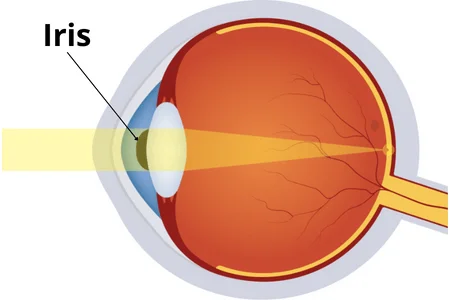
Iris
The iris is the colored part of the eye that surrounds the pupil, controlling the amount of light entering the eye. It consists of muscles that contract or relax, adjusting the size of the pupil in response to changing light levels. In bright conditions, the iris contracts, making the pupil smaller to reduce the amount of light entering the eye. In dim lighting, the iris expands, allowing the pupil to enlarge and gather more light. This automatic regulation helps protect the retina from excessive light exposure and ensures optimal vision in different lighting environments.
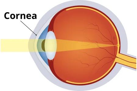
Cornea
The cornea is the transparent, dome-shaped outermost layer of the eye. It plays a crucial role in vision by refracting or bending light as it enters the eye. This bending of light helps focus it onto the retina at the back of the eye, where visual information is processed. The cornea also acts as a protective barrier against dust, germs, and other foreign particles. It lacks blood vessels and receives nutrients from tears and aqueous humor. The cornea’s clarity and shape are vital for clear vision, and any irregularities can lead to vision problems.
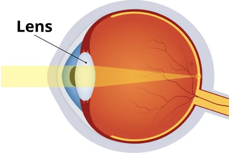
Lens
The lens in the eye is a clear, flexible structure located behind the iris and pupil. It plays a key role in focusing incoming light onto the retina. The lens changes its shape to adjust the focal length, allowing us to see objects at various distances clearly. This process is called accommodation and is crucial for near and far vision. With age, the lens can lose its flexibility, leading to conditions like presbyopia (difficulty focusing on close objects) and cataracts (clouding of the lens), which can impair vision.
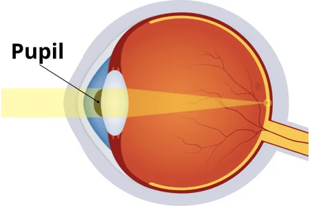
Pupil
The pupil is the dark, central opening in the colored part of the eye called the iris. It regulates the amount of light entering the eye by changing its size. In response to lighting conditions, the iris’s muscles contract or relax, causing the pupil to constrict in bright light, reducing the amount of light entering the eye, and dilate in dim light, allowing more light to enter. This automatic adjustment helps protect the sensitive retina at the back of the eye and ensures optimal vision in varying lighting environments.
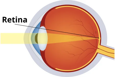
Retina
The retina is a thin layer of tissue at the back of the eye that contains specialized photoreceptor cells called rods and cones. It plays a crucial role in vision by capturing and converting light into electrical signals. Rods are responsible for low-light and peripheral vision, while cones are responsible for color and sharp central vision. These signals are then transmitted via the optic nerve to the brain for processing, allowing us to perceive the images we see. The retina’s health is vital for clear and accurate vision.